
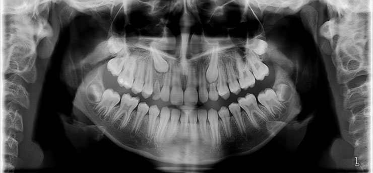
The decision to start orthodontic treatment is a significant step toward upgrading oral health and smile aesthetics. In recent years, the Invisalign method,with its clear, removable aligners, has gained the trust of millions of people worldwide, offering a discreet and comfortable alternative to traditional braces. However, behind the advanced technology of the clear trays and the 3D digital design lies a fundamental stage, without which no treatment can safely begin: a detailed panoramic x-ray.
At the heart of this preparatory phase is the panoramic X-ray. This is a basic imaging examination that captures the entire stomatognathic system in a single image. Although many prospective patients focus primarily on the ClinCheck (the 3D video showing the projected movement of the teeth), this radiographic imaging forms the foundation upon which every successful treatment plan is built.
What is a Panoramic X-Ray and What Does It Reveal to the Specialist?
A panoramic X-ray is a two-dimensional radiographic examination that provides a broad, unified view of the jaws, all the teeth (including those that have not erupted yet), the sinuses, and the temporomandibular joints. Unlike small intraoral X-rays that focus on two or three teeth, this examination offers a comprehensive mapping of the oral cavity with a minimal dose of radiation.
For planning Invisalign treatment, this specific examination reveals critical anatomical details that are impossible to detect through a simple clinical examination or a digital scan. More specifically, it provides information regarding:
-
The length, shape, and direction of the roots of all teeth.
-
The quality and quantity of the alveolar bone surrounding and supporting the roots.
-
The existence of impacted teeth, such as wisdom teeth, or supernumerary (extra) teeth.
-
The presence of intraoral lesions, cysts, inflammation, or even bone resorption.
-
The condition of past endodontic treatments (root canals) and the existence of cavities in non-visible areas.
Knowledge of all these data allows the doctor to evaluate whether the underlying foundation is healthy and ready to accept the forces exerted by the clear aligners to move the teeth.
The Importance of Root and Bone Evaluation Before Aligner Treatment
The orthodontic process relies on a continuous biological response: when controlled pressure is exerted on a tooth, bone on one side resorbs and bone on the other side regenerates, allowing the tooth to shift into its new position. This process directly affects the roots and the periodontium. Therefore, a detailed study of their condition via a panoramic X-ray is of vital importance.
If a patient presents with short roots, the application of orthodontic forces must be performed with extreme caution, using gentle pressure and continuous monitoring, as there is a risk of further root shortening, which could threaten the stability of the tooth. Furthermore, the direction and inclination of the roots determine how the movement must be designed in the ClinCheck software. If the roots of two adjacent teeth converge or are at an unusual angle, moving them blindly without radiographic control could lead to the roots colliding with each other, causing permanent damage or severe pain.
The quality of the bone substrate is the second deciding factor. In cases with a history of periodontitis or periodontal disease, the panoramic X-ray shows the degree of bone loss. If the bone has receded significantly, the treatment plan must be radically modified or postponed until the periodontal condition stabilizes, ensuring that the teeth do not lose their support during Invisalign treatment.
Detecting Hidden Problems with a Panoramic X-Ray
One of the greatest advantages offered by a panoramic X-ray is its ability to bring to light problems that remain hidden beneath the gums. Wisdom teeth (third molars) constitute the most common source of such surprises. Many people have impacted or semi-impacted wisdom teeth that have not erupted into the oral cavity but lie in a horizontal or ectopic position inside the jawbone.
During the planning of an Invisalign treatment, detecting these teeth is pivotal for the following reasons:
-
Space projection: If the treatment requires moving the back teeth backward (distalization) to create space for the front teeth, the presence of an impacted wisdom tooth can completely block this movement.
-
Prevention of inflammation: A semi-impacted wisdom tooth can cause pericoronitis during treatment, forcing the patient to interrupt the use of the aligners, which sets back the entire schedule.
-
Protection of adjacent teeth: Impacted wisdom teeth pressing against the roots of the second molars can cause resorption of the roots of these valuable teeth.
Aside from wisdom teeth, the X-ray can reveal impacted canines or supernumerary teeth, as well as asymptomatic cysts or jaw lesions. Diagnosing these conditions allows the doctor to decide whether an extraction is required before starting Invisalign or if treatment can proceed safely by bypassing or properly incorporating these elements into the plan.
How the Panoramic Examination Influences the ClinCheck Digital Plan
Invisalign technology relies heavily on ClinCheck, a sophisticated 3D imaging software that allows the digital representation of the entire orthodontic journey from the initial stage to the final result. The doctor uses an intraoral scanner to create a precise 3D model of the dental crowns. However, this scan stops at the gumline; it cannot see what is happening underneath.
This is exactly where the panoramic X-ray fills the gap. Designing in ClinCheck cannot be done blindly, relying solely on the aesthetic appearance of the teeth. The doctor integrates the X-ray data into the design to determine the following:
-
Limits of movement: The software can theoretically move a tooth into any position, but in reality, movement is restricted by the available bone. The X-ray shows the actual boundaries within which the root of the tooth can safely move.
-
Placement of Attachments: Attachments are small, protruding elements made of composite resin placed on the teeth to help the aligners exert the necessary forces. Their position directly depends on the axis and inclination of the tooth root—elements that become visible only radiographically.
-
Synchronization of root and crown movements: For a tooth to move correctly, both the visible crown and the root must shift. If the panoramic X-ray shows that a root has a severe inclination, the ClinCheck is programmed so that the forces focus first on correcting the root, ensuring a stable and long-lasting result.
Consequently, this radiographic examination serves as the bridge between the virtual reality of the computer and the biological reality of the patient’s mouth.
Risks and Complications from Bypassing the Panoramic X-Ray
Bypassing the panoramic examination before starting Invisalign treatment constitutes a serious medical error and can pose a direct threat to the patient’s oral health.
Some of the most serious complications that can arise include:
-
Tooth loss due to periodontal destruction: If active, undiagnosed periodontitis is present, orthodontic movement will dramatically accelerate bone destruction, leading to severe tooth mobility or even the need for extraction.
-
Tooth necrosis: If a tooth has suffered past trauma or has deep, non-visible decay/inflammation at the root, the pressure from the aligner can cause acute inflammation, intense pain, and nerve necrosis, requiring immediate root canal treatment and the suspension of Invisalign.
-
Root resorption: As mentioned, moving teeth with already compromised or short roots without specific force modification can lead to complete root resorption, leaving the tooth without support.
-
Treatment failure: The treatment may stop tracking after a few months because the teeth encounter an invisible obstacle inside the bone (e.g., an impacted tooth or a dense bone area), resulting in the aligners no longer fitting properly.
Performing the examination acts as a safety net, protecting the patient from painful situations and unforeseen expenses, while also securing the doctor by ensuring the treatment stands on solid ground.
You might be interested: Malocclusion of teeth: Can it be corrected with Invisalign?
Panoramic X-Ray vs. 3D Scanning and CBCT: How They Complement Invisalign
In modern orthodontics, diagnosis relies on a combination of technologies, where each one fulfills a distinct and irreplaceable role. It is important to understand how the panoramic X-ray collaborates with other digital tools to create the perfect smile.
| Diagnostic Tool | What It Images | Main Role in Invisalign |
| Intraoral Scanning (3D Scan) | The visible part of the teeth (crowns) and the gums with absolute millimeter accuracy. | Used to manufacture the aligners and digitally simulate surface movements. |
| Panoramic X-Ray | A comprehensive, two-dimensional view of the jaws, roots, bone, and impacted elements. | Provides the general health and safety check of the foundation, identifying hidden pathologies and anatomical restrictions. |
| Cone Beam CT (CBCT / Digital Tomography) | A three-dimensional imaging of bone structures and roots with high-definition detail. | Employed selectively in complex cases (e.g., severely impacted teeth) to provide precise spatial awareness. |
The 3D scan shows the orthodontist the starting point and the destination of the outer appearance of the teeth, but the panoramic X-ray is what shows whether the road between these two points is clear and safe. It is not a choice between one tool or another, but rather a comprehensive diagnostic team.
The Role of the Specialized Orthodontist in Treatment Success
Gathering all these diagnostic elements and interpreting them correctly requires deep knowledge and experience, qualities that only an orthodontist specializing in Invisalign can offer. The success of the treatment does not depend simply on the company’s software, but on the ability of the specialist to modify the ClinCheck digital plan based on the findings presented by each patient’s panoramic X-ray.
The Orthodontist in Chalandri and Koropi, Dr. Angeliki Nikolopoulou, can identify subtle anatomical peculiarities, diagnose early any limitations in the bone or roots, and design customized, biologically tolerable movements. In this way, potential complications can be prevented before they appear, complex crowding or alignment issues can be resolved effectively, and the process can be guided with absolute safety, ensuring a healthy, functional, and aesthetically perfect smile that will last over time.